


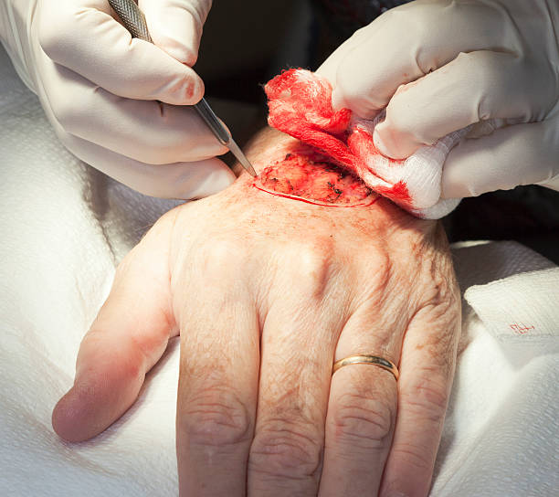
Mohs skin cancer surgery removes cancerous tissue layer by layer. Each layer gets examined under a microscope; the process continues until no cancer remains. The technique targets cancerous cells while preserving as much healthy skin as possible; it provides results during the same appointment. Because the surgeon examines each layer before removing more tissue, the procedure limits the amount of healthy skin removed.
Exploring Mohs Skin Cancer Surgery
Mohs skin cancer surgery treats several types of skin cancer. Basal cell carcinoma and squamous cell carcinoma are the most common types treated with this method; some other skin cancers also qualify. Doctors use this approach for cancers that have a high risk of returning or have returned after earlier treatment. It also suits tumors in areas where preserving tissue matters, including the eyes, ears, nose and hands. When a tumor has unclear edges, is large or grows quickly, Mohs surgery can help the surgeon locate and remove cancerous tissue more precisely.
Removing Cancerous Tissue
The main goal is to remove all cancerous tissue while preserving surrounding healthy skin. The layered method lets the surgeon examine tissue during the procedure; another layer is removed only when cancer remains. This approach can reduce the amount of healthy tissue removed, which is useful when tumors occur in areas where tissue preservation matters. Most patients return home after the procedure; they know whether the examined tissue contains cancer before leaving.
Following the Surgical Steps
A surgeon cleans the treatment area and gives a local anesthetic to numb the skin. The injection can cause brief discomfort, but the treatment area becomes numb before the procedure begins. A surgeon then removes the visible tumor along with a thin layer of tissue around and beneath it; the sample goes to a nearby laboratory for examination.
In the laboratory, the surgeon or a technician cuts the tissue into sections and examines each section under a microscope. A map tracks the location of each piece, so the surgeon knows where cancer remains if it appears in a sample. If cancer is present, another thin layer is removed from that specific area; the patient waits while the new sample is examined. The cycle continues until the tissue sample shows no remaining cancer.
Repairing the Wound
After all cancer has been removed, the surgeon determines how to repair the wound. The method depends on the wound’s size and location, and most repairs take place during the same appointment. Common options include:
- Allowing the wound to heal naturally
- Closing the wound with stitches
- Moving nearby skin over the wound
- Using a skin graft from another part of the body
Large or complex wounds sometimes require temporary closure. A separate repair procedure may be needed in some cases, and the surgeon explains the available options after removing the cancer. Mild pain and soreness can occur after the procedure. Over-the-counter pain relievers can usually manage this discomfort, while severe pain is uncommon. Patients wear a bandage for a week or longer, and they receive instructions for cleaning the site and changing the dressing. Strenuous exercise and heavy lifting should be avoided for at least one week, and patients follow their wound-care instructions during recovery. Follow-up appointments help monitor healing, while prescribed ointments may be used as directed.
Schedule a Consultation
A consultation allows a dermatologist to evaluate the skin cancer and discuss whether Mohs skin cancer surgery fits the treatment plan. The care team reviews what to expect before, during and after the procedure, and patients receive instructions about medications and other preparation steps. Vivida recommends stopping certain blood-thinning medications and supplements one to two weeks before surgery, while Mayo Clinic advises patients to follow their surgeon’s specific medication instructions. The surgeon can also explain wound repair and recovery based on the procedure.
